OBSERVATION · SKIN & COAT
How should I record my pet’s skin and coat changes for the veterinarian?
Use consistent photos, a body location and a short timeline so the clinic can see what changed—and what remains unknown.
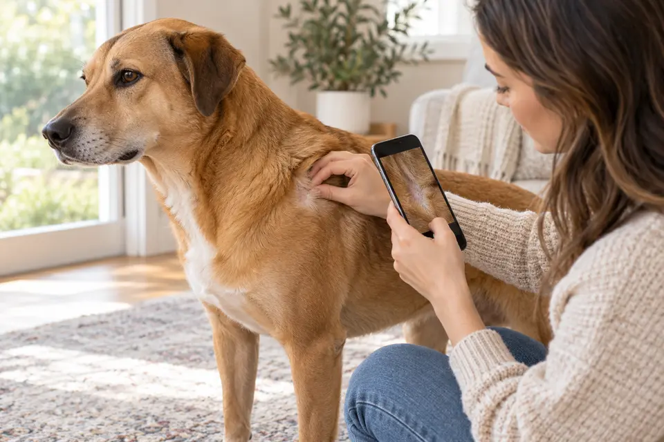
THE DIRECT ANSWER
Record when the change was first noticed, where it is, how widely it appears, whether it has moved or changed, and what licking, scratching or rubbing you actually saw. Take a well-lit overview and a closer photo from repeatable angles without cleaning, squeezing or treating the area first unless a veterinarian directs you. Include recent husbandry, travel, animal contact and product changes. A photograph can document appearance and progression; it cannot identify the cause or replace a full examination.
Sources: Merck Veterinary Manual: Diagnosis of Skin Diseases in Small Animals · Cornell Animal Health Diagnostic Center: Dermatopathology
Make the location repeatable
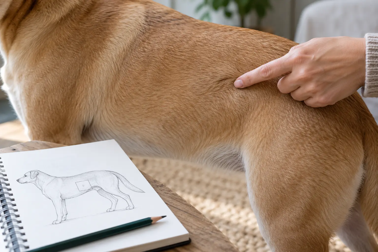
Merck explains that dermatologic history and lesion distribution help a veterinarian interpret the physical examination and choose tests. Use ordinary body landmarks—left shoulder, underside of abdomen, between right-front toes—rather than a diagnosis word.
Begin with one whole-body or regional photo, then a closer view. Use similar lighting and angle on later dates. A coin, ruler or color chart can mislead if perspective changes; if the clinic wants scale, ask what method it prefers.
Do not crop away the surrounding coat or digitally enhance color. Keep the original date and note whether fur was parted, wet or recently groomed.
| Field | Useful fact | Do not turn it into |
|---|---|---|
| Location | Side, body landmark and area involved | A disease label |
| Time | First noticed and dates rechecked | A guessed onset when nobody saw it |
| Appearance | Color, hair, surface and moisture in plain words | A cause inferred from a photo |
| Behavior | Observed licking, scratching or rubbing | An itch score invented after the event |
| Context | Products, grooming, travel, animal contact and routine changes | Proof that one exposure caused it |
Sources: Merck Veterinary Manual: Diagnosis of Skin Diseases in Small Animals · Cornell Animal Health Diagnostic Center: Dermatopathology
Keep appearance, behavior and context in separate lines
A bare-looking patch may follow self-trauma, coat breakage, clipping or several other processes. Record what is visible and what the pet did without joining them with 'because.'
Include normal findings that bound the record: the matching area on the other side looked unchanged, appetite was usual, or no licking was seen during a specific window. 'Not seen' is not the same as 'did not happen.'
If a veterinarian may sample the area, ask before washing, applying a topical product or removing crusts. Merck notes that surface material can matter to some diagnostic procedures.
Sources: Merck Veterinary Manual: Diagnosis of Skin Diseases in Small Animals
A fictional three-day record
Fictional example: Noor notices a small thin-coated area over dog Pippin's left shoulder on Monday evening. She takes a regional and close photo in window light, then records two brief licking episodes she personally sees.
On Wednesday the area looks wider in a repeat-angle photo, but the Tuesday daytime period was unobserved. Noor records a new shampoo used the previous weekend without calling it the cause and contacts the clinic.
The example is a documentation workflow, not a patient case. No diagnosis, treatment or safe waiting period follows from these observations.
| Date | Observed | Unknown |
|---|---|---|
| Monday | Thin-coated area at left shoulder; two witnessed licking episodes | Exact onset and cause |
| Tuesday | No evening photo change obvious | Daytime behavior was not observed |
| Wednesday | Repeat-angle photo appears wider; clinic contacted | Whether shampoo or another factor is relevant |
Bring originals and a short summary
Cornell's dermatopathology service asks for a complete clinical and dermatologic history, lesion location and digital images. That laboratory guidance shows why a photo is useful beside history; it does not mean an owner can classify a lesion from the image.
Keep the best two or three original images available rather than sending a long unlabeled camera roll. Add the pet, location, date, progression and any products already used.
Ask the clinic how it wants images delivered. My Pet Notes can hold a local text summary, but it does not upload photos to a clinic or examine skin.
Sources: Cornell Animal Health Diagnostic Center: Dermatopathology
Choose the next observation without delaying care
If the clinic advises a recheck, write the date, lighting and angle you will repeat. Do not wait for a planned comparison when urgent signs appear.
Keep medication, parasite-product and grooming records exact. A brand family or 'anti-itch cream' is not enough for a clinician to identify an exposure.
The goal is a usable handoff: what changed, where, when, and what is still uncertain.
- Location and side are unambiguous.
- Photo dates and observation gaps remain visible.
- Behavior and appearance are recorded separately.
- Products and recent context use exact names where known.
FROM THE GUIDE · INTO THEIR EVERYDAY
A skin-and-coat observation
Keep location, photo context and observed behavior separate. This does not diagnose a skin condition.
Draft my skin observationChoose their notebook, add your own details, then save. Free, no account. Notes stay in this browser.
THE NEXT QUESTIONS
What else might you be wondering?
Can a clear photo tell my veterinarian what the skin problem is?
It can preserve appearance, location and change over time, but diagnosis requires the history, physical examination and sometimes testing.
Sources: Merck Veterinary Manual: Diagnosis of Skin Diseases in Small Animals · Cornell Animal Health Diagnostic Center: Dermatopathology
Should I wash the area before the appointment?
Ask the clinic. Cleaning or applying a product can alter surface material that may matter to examination or sampling.
Sources: Merck Veterinary Manual: Diagnosis of Skin Diseases in Small Animals
What if I never saw my pet scratch?
Record the window you observed and say scratching was not seen then. Do not convert an unobserved period into proof that itching never occurred.
LOOK CLOSER AT THE EVIDENCE
Sources & what they establish
Official guidance, research findings and PetCheck’s practical interpretations have different roles. These notes explain how the sources were used.
Veterinary reference
Merck dermatologic history and examination
Supports recording distribution, region, history and wider health context.
Limit: Clinical lesion terms and diagnostic tests belong to veterinary examination, not photo matching.
Sources: Merck Veterinary Manual: Diagnosis of Skin Diseases in Small Animals
University laboratory guidance
Cornell dermatopathology submissions
Supports location, history and digital images as useful supporting material.
Limit: Submission guidance does not validate an owner's diagnosis or image interpretation.
Sources: Cornell Animal Health Diagnostic Center: Dermatopathology
Original editorial workflow
Pippin's fictional timeline
Shows how to preserve progression, exposure and an observation gap.
Limit: Not a real patient, diagnosis or treatment outcome.
Sources checked 2026-10-22. Examples are fictional and illustrate reasoning, not patient outcomes. This is an editorial source review, not independent veterinary review. How we prepare these guides →
FOR ANOTHER VISIT
Keep this guide close.
Save it to read again, or copy a link to share.
ONE QUESTION CAN LEAD TO ANOTHER
A useful next read.
Choose the question that fits what you want to understand next.
 AI-generated editorial scene
What should I write down before my pet’s vet visit?
Place the skin-and-coat record inside the wider clinic timeline without adding a diagnosis.
Read the guide AI-generated editorial scene
How do I use flea and tick products safely around my pets?
Keep any recent parasite-product name and application record exact and separate.
Read the guide